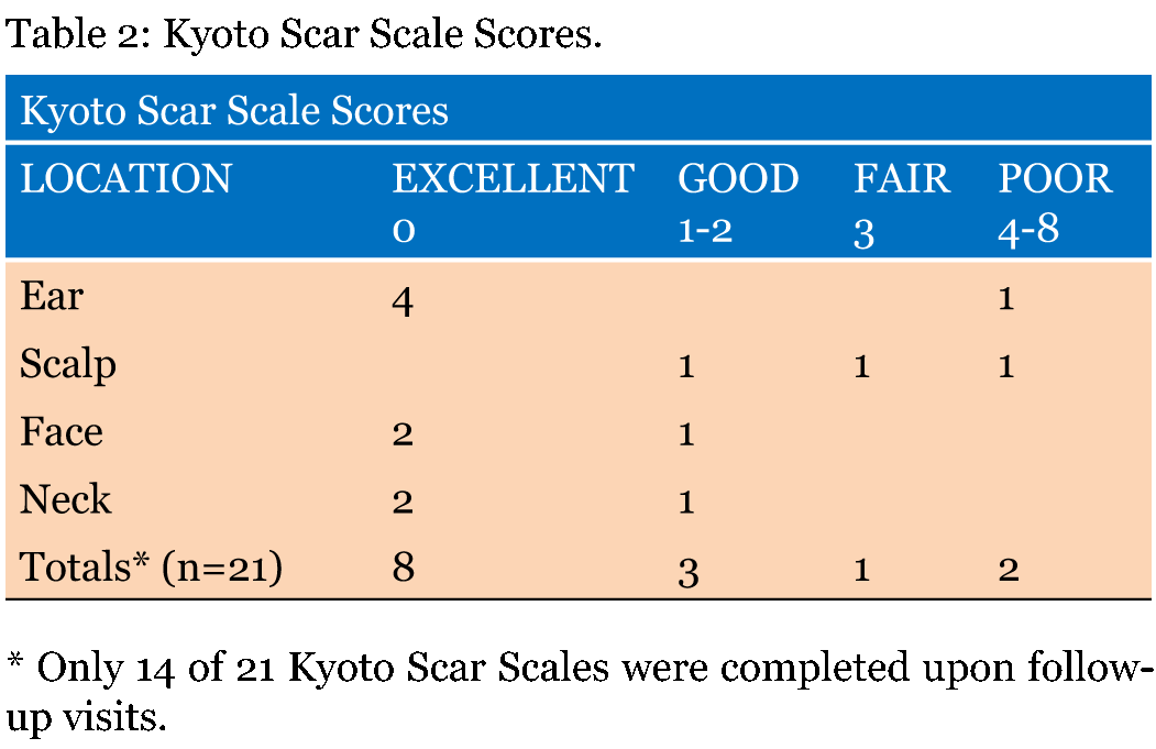
| Table of Contents | |
|
Original Article
| ||||||
| Head and neck keloid management: A retrospective early review on a new approach using surgical excision, platelet rich plasma and in office superficial photon X-ray radiation | ||||||
| Michael Eugene Jones1, Cherrell Jackee Hardy2, Julie Marie Ridgway3 | ||||||
|
1MD, Founder/Medical Director, Lexington Plastic Surgeons, New York, USA.
2RN, BSN, Nurse Manager, Lexington Plastic Surgeons, New York, USA. 3RN, BSN, Registered Nurse, Lexington Plastic Surgeons, New York, USA. | ||||||
| ||||||
|
[HTML Abstract]
[PDF Full Text]
[Print This Article]
[Similar article in Pumed] [Similar article in Google Scholar] |
| How to cite this article |
| Jones ME, Hardy CJ, Ridgway JM. Head and neck keloid management: A retrospective early review on a new approach using surgical excision, platelet rich plasma and in-office superficial photon X-ray radiation. Edorium J Otolaryngol 2015;2:14–19. |
|
Abstract
|
|
Introduction:
The objective of this retrospective study was to evaluate the efficacy of our combination therapy protocol for head and neck keloid treatment.
Methods: We treated keloids using surgical excision, platelet-rich plasma and post-operative in office superficial photon X-ray radiation therapy. Results: No recurrences determined. Two of 21 keloids displayed poor results. Conclusion: Surgical excision combined with platelet rich plasma and postoperative in office superficial radiation therapy achieved a 100% non-recurrence rate at 4th to 11th month follow-up. This protocol appears to be a viable alternative in the management of keloids. | |
|
Keywords:
Head, Keloid, Neck radiation, Surgery, Triamcinolone
| |
|
Introduction
| ||||||
|
Keloids are fibrous lesions that form at a site of injury due to irregular production of type III and type I collagen [1] . Unlike hypertrophic scars, keloids continue to grow outside of the original wound margins, fail to resolve over time, may itch and become painful [2]. High tension areas such as the chest, trunk and back, as well as commonly pierced areas such as the ears, are some of the more popular sites for keloid formation [3] [4]. While there is limited understanding on the cause of keloids, they are known to occur more frequently in persons age 10–30, and in females (likely the result of more frequent body piercing) [1]. Although documented in all races, keloids are more prevalent in people of color (African American, Asian, Latinos) with a positive correlation to skin pigmentation. Most keloids appear in those with darker skin types while there are no reported incidents in Albinos [2]. Keloids often mature into unsightly lesions affecting self-esteem and quality of life. Depending on the location and size of the keloid, range of motion may be impaired [4]. With limited knowledge about the actual cause of keloids, determining the most effective treatment for keloids has proven challenging. It is agreed, however, that combination therapies are necessary to reach lower recurrence rates. No single therapeutic modality alone has proven effective. Treatments such as surgical excision, postoperative radiotherapy, triamcinolone injections, pulse dye laser, cryotherapy and silicone sheeting are amongst the more popular treatments used [4]. Many combinations of these therapies have been used with results varying drastically. In some studies, surgical excision alone results in a recurrence rate of 65–99% [5]. Other studies reported surgical excision combined with other adjuvant therapies such as cryotherapy, pressure treatment, intralesional steroids and silicone gel sheeting had a recurrence rate greater than 50%, while surgical excision followed by radiation therapy proved most effect with a recurrence rate of nearly 20% [6]. Multimodality therapy involving surgical excision, postoperative radiotherapy and intralesional steroid injections yielded up to 89% cure rate in one study [7]. Although not studied in the treatment of keloids, platelet rich plasma (PRP) has shown favorable wound healing properties demonstrating organized collagen deposition [8]. As a result, patients were treated using surgical excision, PRP, and in office superficial photon radiation therapy. | ||||||
|
Materials and Methods
| ||||||
|
From November 2013 to June 2014 at Lexington Plastic Surgeons in New York, we treated keloids with extralesional surgical excision, platelet rich plasma (PRP) and postoperative superficial radiation therapy. A total of 21 keloids were treated in 20 patients. Patients in this study were assessed at a minimum postoperative period of four months and a maximum period of 11 months. Cumulative radiation doses ranged from 13 Gy to 18 Gy. Of the 21 keloids 71% belonged to African Americans, 57% of keloids were in females, and the ear was the most common treatment area making up 11/21 (52%) of lesions treated (Table 1). Surgical Procedure Radiation Protocol From November 2013 to February 2014 eleven keloids were treated using two fractions for a cumulative radiation dose of approximately 16 Gy. With the delivery of two fractions and 16 Gy, it was noted that patients developed significant hyperpigmentation in the treatment field. To reduce the post inflammatory response and hyperpigmentation, treatments were increased to three fractions. From April 2014 to June 2014 nine keloids were treated using three fractions for a cumulative radiation dose of approximately 18 Gy. Due to personal limitations the remaining keloid was treated using one fraction for a cumulative radiation dose of approximately 13 Gy. Clinical Follow-Up Assessment | ||||||
| ||||||
|
| ||||||
| ||||||
|
Results | ||||||
|
From November 2013 to October 2014, 20 patients (21 keloids) were treated and followed from 4 to 11 months. Treatment of the keloids included extralesional surgical excision with intraoperative platelet rich plasma followed by post-operative in office superficial photon X-ray radiation therapy (mean cumulative dose 1682.4 cGy over 2–3 days). Of the 21 keloid lesions, nine occurred in men and twelve in women with a mean age of 36.5 (range 16–66). During the follow-up period, there was no evidence of recurrence as defined by any sign of extraordinary erythema, induration and hypertrophy of the scar beyond the site of excision. Thus, there was a 100% non-recurrence rate at four to eleven month follow-up. There were only two lesions (9.5%) that were qualified as poor by the Kyoto scale, due to mild hypertrophy and warranted triamcinolone 10 mg injections. Radiation induced hyperpigmentation was noted in all patients. However, hyperpigmentation improved when treatment increased from two to three fractions of radiation. Fourteen postoperative scars were evaluated by the Kyoto scar scale: 57% (excellent), 21% (good), 7% (fair), 14% (poor) (Table 2). | ||||||
|
Discussion
| ||||||
|
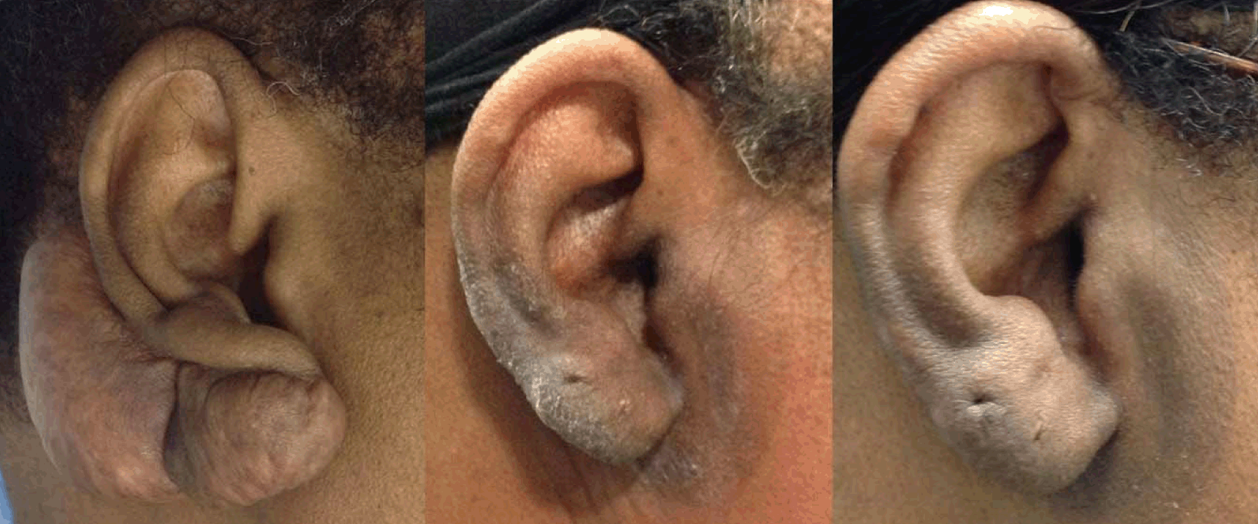
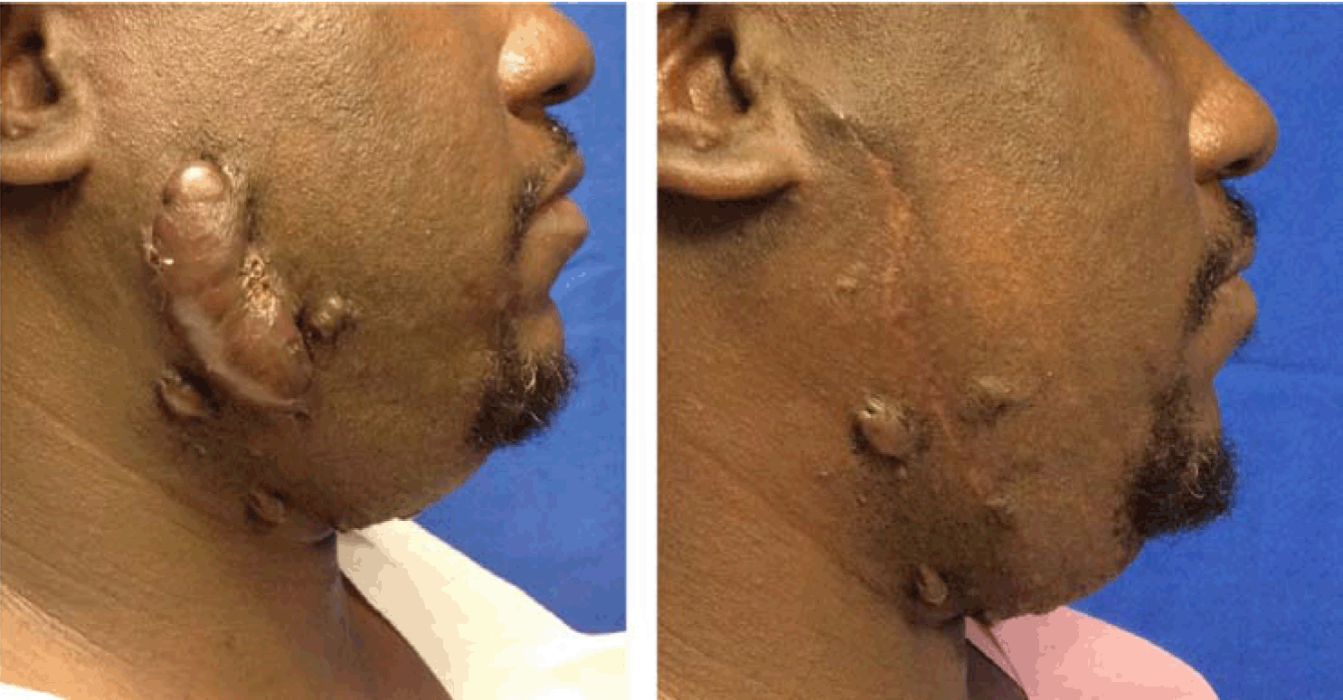
In this study, we present a new and innovative in-office treatment protocol using surgical excision, platelet rich plasma and superficial photon X-ray radiation therapy for the treatment of keloids. Twenty-one keloids were treated with this modality. In this early retrospective case review, we have observed a non-recurrence rate of 100%. Keloids are benign yet locally invasive lesions related to complex cellular, environmental and genetic interactions [5]. These lesions are more common amongst people of darker skin types (Asians, Latinos, Jews and people of African descent) [9]. Although there is a multitude of treatment modalities for keloids, none are satisfactory. The use of silicone sheets, cryotherapy, laser treatments, and pressure dressings are among the conservative treatments recommended, but the effect of these modalities is limited. The use of intralesional steroid injections, either prior to or after surgical excision, has yielded less than optimal results. Multimodality therapy involving surgical excision, postoperative radiotherapy and intralesional steroid injections has yielded up to 89% cure rate in one study [7]. Some studies even report surgical excision, triamcinolone injections and radiotherapy to yield less than 10% recurrence rate [10]. In this study, we present a new and innovative in-office treatment protocol using surgical excision, platelet rich plasma and superficial photon X-ray radiation therapy for the treatment of keloids. In this early retrospective case review, we have observed a non-recurrence rate of 100%. Radiation: This is the first study to our knowledge to report on the use of in-office superficial photon X-ray radiation in the treatment of keloids. This low energy therapy may be advantageous over more traditional radiation machines that utilize high-energy electron beams. The device focuses the photon X-rays into the skin only, where the pathology is located and does not penetrate and impact the underlying tissue [11]. Due to the rapid fall off of radiation, exposure to surrounding tissues is limited [11]. Electron beam radiotherapy has the ability to penetrate beyond the surface of the skin missing the pathology and impacting underlying structures [11]. Further, the superficial radiation therapy was easier for us to coordinate with the surgical excision, since both occurred in the same office. In the treatment of keloids, irradiation dose, fraction and period from surgical excision to irradiation are important. There currently is no consensus on the ideal radiation dose to be administered. Fifteen to 20 Gy has been used in many protocols [7]. In our study, patients were treated with 13–18 Gy over one to three fractions with a biological effective dose (BED) of 30 Gy [10]. It is commonly accepted that radiation treatment should be started within 72 hours of the surgical excision [13]. In our study, all patients were treated within 72 hours of surgical excision. Recurrence: For the purpose of this study recurrence was defined as any sign of extraordinary erythema, induration and hypertrophy beyond the site of excision. No keloids qualified as recurrence. Post irradiation hyperpigmentation: Most patients reported overall satisfaction with their treatment outcomes. Post irradiation hyperpigmentation was the most disliked side effect of radiotherapy. Since high dose radiation has been reported to damage melanocytes and decrease hyperpigmentation, our protocol was changed from two to three fractions [13]. The increase in fractions allowed for the delivery of high dose radiation. Patients were also allowed to bathe with mild cleanser and water and use topical steroids which have been proven to lessen the radiation induced skin reactions [14] [15]. After the change of protocol a marked difference was noted in the severity of hyperpigmentation in the radiation field (Figure 1). Triamcinolone: Triamcinolone is widely used in the treatment of keloids and hypertrophic scars. Studies have shown decreased keloid fibroblast proliferation, degradation of mature collagen and enhanced organization of existing collagen bundles in lesions treated with triamcinolone [5]. All three findings are critical in the treatment of keloids and hypertrophic scars. Intralesional triamcinolone 10 mg injections were administered to two lesions with poor results by the Kyoto scale due to hypertrophy. One injection was performed for each and subsequent follow-up demonstrated no further progression of the hypertrophy at the time of the manuscript. Ongoing monitoring will continue. Platelet Rich Plasma: Platelet Rich Plasma (PRP) is autologous platelet concentrate shown to stimulate tissue repair [16]. In addition to accelerating wound healing, PRP has been shown to decrease pain and disability in the treated area [8] [17]. Further, research has shown decreased inflammation and collagen deposition, as well as more organized collagen structures in injured vocal cords treated with PRP [18]. In our practice, evidence of improved wound healing with lower keloid recurrence was noted anecdotally with surgical excision and PRP alone, prior to the use of in office radiation therapy. As a result, all patients received PRP to the surgical site. Kyoto Scar Scale: The Kyoto Scar scale was used to qualify scars during postoperative visits. As pain and itching are expected after surgery the scores were negatively impacted in the early phases of wound healing. Of the 21 keloids treated (14 completed the assessment) eight qualified as excellent (Figure 2) and (Figure 3), three were good, one was fair and two were poor. The most common qualifying symptom was partial hardness. Normal early post-surgical inflammation can contribute to hardness or induration along the incision. Signs of recurrence will be closely monitored. Short Follow-Up: The short follow-up period is a limitation of this study. At maximum patients were eleven months post-surgical excision and radiotherapy. Keloids are widely considered cured after 3–5 years without recurrence. Ongoing monitoring will continue. | ||||||
|
| ||||||
|
| ||||||
|
| ||||||
|
| ||||||
|
Conclusion
| ||||||
|
Surgical excision combined with postoperative in office superficial radiation therapy achieved a preliminary 100% non-recurrence rate at four to eleven month follow-up. Hyperpigmentation was noted in all patients. The study was limited by the short follow-up period, the non-standardized radiation protocol, variable use of triamcinolone and inconsistent use of the Kyoto scar scale. Early results of this study demonstrate that this protocol appears to be a safe and viable option in the management of keloids and merits further randomized controlled study of its comparative efficacy. | ||||||
|
References
| ||||||
| ||||||
|
[HTML Abstract]
[PDF Full Text]
|
|
Author Contributions:
Michael Eugene Jones – Substantial contributions to conception and design, Acquisition of data, Analysis and interpretation of data, Drafting the article, Revising it critically for important intellectual content, Final approval of the version to be published Cherrell Jackee Hardy – Analysis and interpretation of data, Revising it critically for important intellectual content, Final approval of the version to be published Julie Marie Ridgway – Analysis and interpretation of data, Revising it critically for important intellectual content, Final approval of the version to be published |
|
Guarantor of submission
The corresponding author is the guarantor of submission. |
|
Source of support
None |
|
Conflict of interest
Authors declare no conflict of interest. |
|
Copyright
© 2015 Michael Eugene Jones et al. This article is distributed under the terms of Creative Commons Attribution License which permits unrestricted use, distribution and reproduction in any medium provided the original author(s) and original publisher are properly credited. Please see the copyright policy on the journal website for more information. |
|
|